For decades, taking dental impressions was one of the most uncomfortable parts of a dental visit. Patients dreaded the heavy, gag inducing impression putty, while clinicians spent valuable chairside time battling material bubbles, tears, and physical model distortions.
Enter the Intraoral Scanner (IOS)- The cornerstone of the modern digital dental practice. By replacing physical trays with an optical 3D scanning wand, dental practices are elevating precision, saving time, and dramatically improving patient comfort.
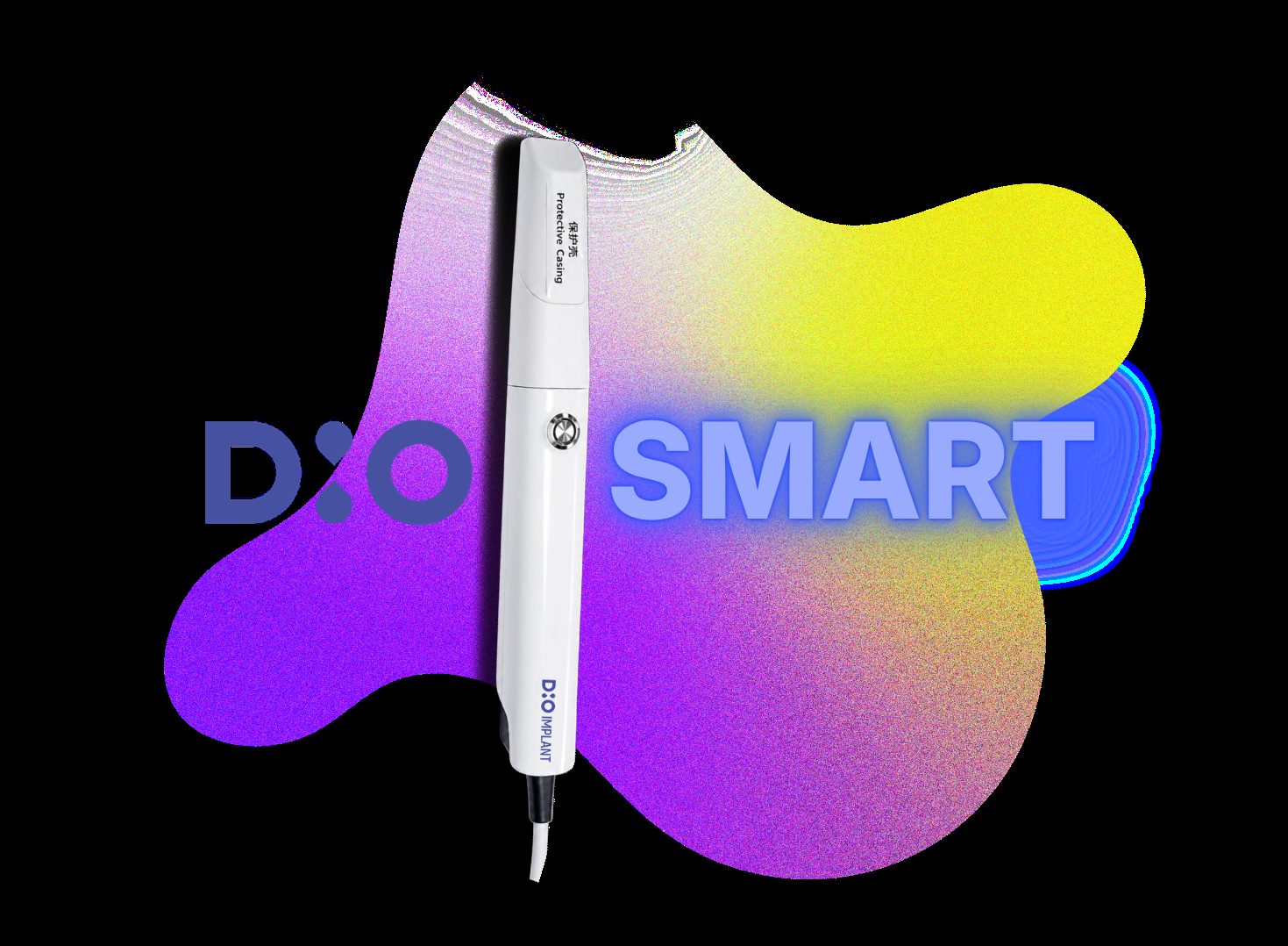
An intraoral scanner is a handheld, ergonomic device that projects a light source (optical laser or LED) onto the surfaces of the teeth and gingival tissues.
As the clinician glides the wand over the dental arch, built-in sensors capture thousands of 3D data points per second. Advanced software processes these images in real time, assembling a hyper accurate, high-definition 3D color model (usually in STL or PLY file formats) right on your monitor screen.
Unmatched Clinical Accuracy: Modern optical scanners capture microscopic margins with high fidelity, eliminating material shrinkage, tearing, and bubble distortions common with traditional putty.
Superior Patient Comfort: Eliminating heavy impression putty creates a fast, gag-free, and comfortable experience that patients appreciate.
Instant Lab Collaboration: Instead of waiting days for courier shipping, digital impression files can be emailed directly to the dental lab within seconds for immediate production.
Interactive Patient Education: Displaying high-definition, 3D color renders of a patient's mouth on screen makes treatment proposals clear and significantly increases case acceptance.
An intraoral scanner isn't just for single crowns, it serves as the digital bridge for complex procedures. When merged with Cone Beam Computed Tomography (CBCT) bone data, intraoral scans allow clinicians to design custom surgical guides for flapless, highly accurate implant placements.
Q1: Does taking a digital scan with an intraoral scanner expose the patient to radiation?
No, intraoral scanners use harmless optical light or laser technology, making the process 100% radiation-free.
Q2: How fast is a typical full-arch intraoral scan?
A skilled practitioner can complete a full arch digital scan in under 1-2 minutes.
Q3: Can intraoral scanners completely replace messy impression putty?
Yes, they fully replace physical impression materials for crowns, aligners, nightguards, and implant restorative planning.
Q4: How do intraoral scanners improve restorative accuracy?
They eliminate material shrinkage, tearing, and bubble distortions common with traditional putty impressions.
Q5: Can intraoral scanner files be shared directly with dental labs?
Yes, digital files (such as STL or PLY formats) can be emailed to lab technicians instantly for rapid fabrication.
Dental Surgery Instruments required for different types of dental implants—endosteal trays, immediate cases, guided kits and full-arch drivers.
Dental Implant Cost In India explained: what quotes should include, pricing factors, quality versus lowest fee, and questions to ask before treatment.
How dental bone graft material and a dental implant motor (physio dispenser) work together for cooler osteotomies and stable grafted implant sites.